| Description |
: |
The organization and cell-lineage of the ascidian egg / by Edwin G. Conklin.
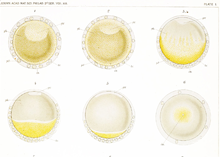
Fig. 1. Unfertilized egg before the fading of the germinal vehicle, showing central mass of gray yolk, peripheral layer of yellow protoplasm, test cells and chorion.
Fig. 2. .Similar egg after the disappearance of the nuclear membrane, showing the spreading of the clear protoplasm of the germinal vesicle at the animal pole.
Fig. 3. Another egg about five minutes after fertilization, showing the streaming of the peripheral protoplasm to the lower pole where the spermatozoon enters, thus exposing the gray yolk of the upper hemisphere; the test cells are also carried by this streaming to the lower hemisphere.
Figs. 4 and 5. Other eggs -bowing successive stages in the collection of the yellow and clear protoplasm at the vegetal pole; clear protoplasm lies beneath and extends a short distance beyond the edge of the yellow cap.
Figs. 6-10. Successive stages of the same egg drawn at intervals of about five minutes; viewed from the vegetal pole. In fig. 6 the area of yellow protoplasm is smallest, and the sperm nucleus is a small clear area near its center. Figs. 7.-10 show stages in the spreading of this yellow protoplasm until it covers nearly the whole of the lower hemisphere; at the same time the sperm nucleus and aster move toward one side of the yellow cap and the yellow protoplasm begins to collect into a crescent at this side.
Fig. 11. Side view of an egg of about the same stage as fig. 10, showing the eccentric position of the sperm nucleus and a small area of clear protoplasm at the upper pole where the polar bodies are being formed.
Fig. 12. Polyspermic (?) egg, viewed from the vegetal pole, showing four collections of yellow protoplasm around as many sperm (?) nuclei (see p. 24). |


