Multi-dimensional marine organisms dataviewer
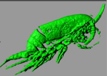
Auto-fluorescence of the Cératiums and the Copepods and reconstruction in 3 dimensions with the Imaris software
| Organism | : | Crustacean |
|---|---|---|
| Species | : | Copepod |
| Description | : | (A1) Corycaeus tipicus copepod. Confocal microscope(10X). Auto-fluorescence. Left image (Ex 405 nm / Em 420-583 nm). Right image(Ex 543 nm / Em 590-700 nm). Scales 100 μm. (A2) Copepod. Confocal microscope(10X). Auto-fluorescence. Left Image(Ex 405 nm / Em 420-583 nm). Right image (Ex 543 nm / Em 590-700 nm). Scales 100 μm. (A3) Copepod. SPIM microscope(10X). Auto-fluorescence(Ex 488 nm / Em 505-545 nm) and 3D construction with Imaris software. (A4) Copepod. Confocal microscope(10X). Auto-fluorescence(Ex 405 nm / Em 420-583 nm) and 3D construction. (A5) Copepod. SPIM microscope(10X). Auto-fluorescence(Ex 488 nm / Em 505-545 nm) and 3D construction. (A6) Appendices of Copepods. Confocal microscope(10X). Auto-fluorescence(Ex 405 nm / Em 420-583 nm) and 3D construction. (A7) Ceratium. Confocal microscope(25X). Auto-fluorescence(Ex 410 nm / Em 535-685 nm) and 3D construction. (A8) Ceratium. Confocal microscope(10X). Auto-fluorescence(Ex 405 nm / Em 420-583 nm). (B1) Ceratium. Epi-fluorescence microscope(10X). Auto-fluorescence. left Image(Ex 488 nm / Em 500-500 nm). Right image (Ex 543 nm / Em LP 590nm). (B2) Ceratium. Epi-fluorescence microscope(10X). Marking Cellmask Green. Left image (Ex 488 nm / Em 500-555 nm). Right image (Ex 543 nm / Em LP 590nm). Scale 100 μm. |
| Attributions | : | Ben aicha Sameh |
| Website | : | http://biodev.obs-vlfr.fr/fr/equipes_de_recherche/fecondation_et_controle_du_cycle_meiotique.html |
| Licensing | : |

|
Click to visualize