Items count : 117

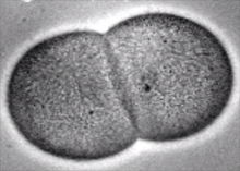
Embryonic development of Phallusia mammillata from 2 cell to gastrulation

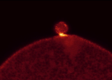
Supplementary Movie 8. Cortical actin patch forms under PB1

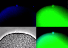
Supplementary Movie 7. DNA can polarize the egg cortex in ascidians as in mouse oocytes.

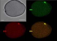
Supplementary Movie 6. Failed rotation of second meiotic spindle giving two second polar bodies

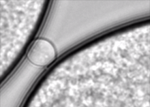
Supplementary Movie 5. Two second polar bodies


Supplementary Movie 4. Second meiotic spindle tilts during emission of PB2

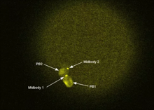
Supplementary Movie 3. Midbody 1 and midbody 1 location between polar bodies

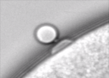
Supplementary Movie 1. Bright field movie showing location of protrusion between PB1 and PB2

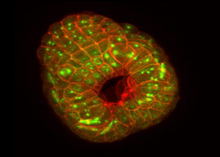
Phallusia development from neural to tadpole

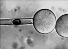
Nocodazole pipette to depolymerise CAB proximal microtubules in zygote

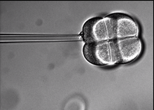
Nocodazole pipette to depolymerize microtubules at the 8-cell stage.

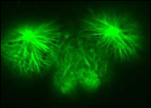
Microtubule growth around the CAB in an intact embryo.

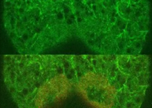
Microtubule polymerization around the CAB.

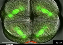
Kif2 protein leaves the CAB during mitosis.

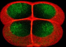
CAB visualization

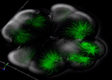
3D rendering of microtubules during UCD

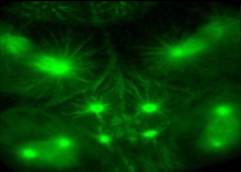
Unequal Cell Division 3. 32 to 64-cell

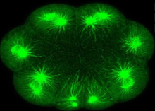
Unequal Cell Division 2. 16 to 32-cell

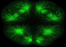
Unequal Cell Division 1. 8-16-cell

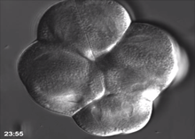
Unequal cell division from 8 to 16-cell stage.

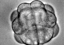
Embryonic development of Phallusia mammillata from 2 cell to gastrulation

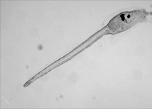
Phallusia mammillata metamorphosis

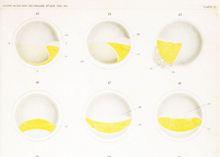
Plate II - Conklin 1905 - First Cleavage

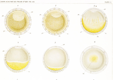
Plate I - Conklin 1905 - Maturation and Fertilization

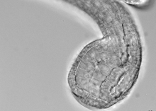
Notochord intercalation

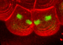
B5.2 cell division in 3D

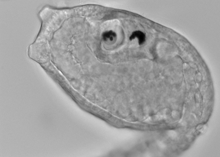
Live-palps

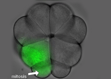
B-catenin speeds up the cell cycle in the ascidian blastula

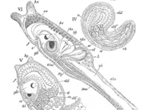
Various stages in the development of Phallusia mammillata


Interkinetic nuclear migration in early ascidian embryos

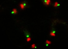
Live centrosome movements in a Phallusia embryo

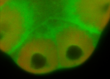
Second Unequal Cleavage of PGC precursors

